


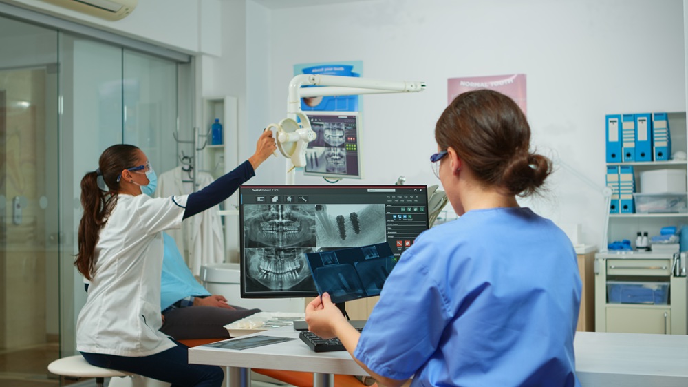
Root canal treatment can sound intimidating, but modern imaging helps dentists understand what is happening inside and around a tooth before treatment begins. If you visit a dental clinic in Hamilton with tooth pain, X-rays may be part of the examination to identify the cause and assess the tooth.
Why Imaging Matters in Root Canal Treatment
A tooth contains soft tissue called the pulp, which includes nerves and blood vessels. When the pulp becomes inflamed or infected, root canal treatment may be needed to remove the affected tissue, clean the canals, and seal the tooth.
Imaging can help your dentist examine the roots, surrounding bone, and areas that may not be visible during a regular examination. If you are considering root canal treatment near you, X-rays and 3D imaging can provide important information for diagnosis and treatment planning.
What Can Dental X-Rays Show?
Traditional dental X-rays remain an important part of root canal diagnosis and treatment planning. Depending on the image taken, they may help show:
- Signs of infection: Changes in the bone around a root tip may suggest infection or inflammation.
- Root anatomy: X-rays can provide information about the number, shape, and length of the roots.
- Previous dental work: X-rays can show fillings, crowns, and previous root canal treatment.
- Bone changes: Bone loss or other changes around the tooth may be visible.
Because standard X-rays create two-dimensional images, some structures may overlap or remain difficult to see.
When Is 3D Imaging Helpful?
Cone beam computed tomography, or CBCT, creates three-dimensional images of teeth, roots, bone, and nearby structures. It can provide extra information when a regular X-ray does not answer a specific diagnostic question.
CBCT may be recommended for more complex situations, such as:
- Unusual anatomy: A tooth may have extra or unusually curved canals.
- Previous treatment: More detail may be useful when assessing a tooth that has already had root canal treatment.
- Suspected complications: CBCT may help investigate some fractures, resorption, or persistent infection.
- Nearby structures: A 3D view can show the relationship between a tooth and surrounding anatomy.
CBCT is not needed for every root canal. The decision depends on the clinical examination and whether the extra information may change diagnosis or treatment planning.
What to Expect During Dental Imaging
Standard dental X-rays are taken with a sensor or image receptor positioned near the tooth. For a CBCT scan, you sit or stand still while the scanner moves around your head.
When you visit a dental clinic near you, the dentist reviews the images together with your symptoms and clinical examination. Imaging does not replace an examination; it provides additional information that can help confirm a diagnosis and plan treatment.
Seeing the Full Picture Before Treatment Begins
The right imaging can help clarify what is happening inside a painful or damaged tooth and guide the next step in care. At Centre Mall Dental, we can assess your symptoms, review your dental health, and explain whether root canal treatment may be appropriate. Contact us today to schedule an appointment.
Frequently Asked Questions
Are dental X-rays safe?
Dental X-rays use ionizing radiation, so they should be taken only when clinically needed. The radiation dose from routine dental X-rays is generally low.
Will I need a 3D scan for every root canal?
No. CBCT is generally reserved for cases where three-dimensional information may help answer a specific diagnostic or treatment-planning question.
Can CBCT detect a cracked tooth?
CBCT can sometimes provide useful information when a fracture is suspected, but not every crack can be seen on a scan.
Does a CBCT scan hurt?
No. The scan itself is non-invasive. You simply remain still while the machine captures the images.